State of the Art
Below are details of our OCT Health Scan and Digital Retinal Photography
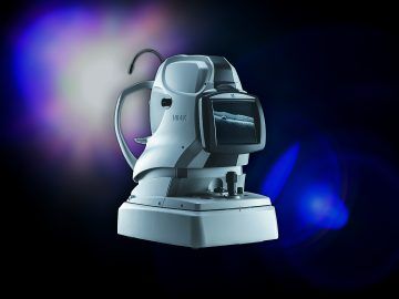
Optical Coherance Tomography (OCT)
Accurate frame and eye measurements are essential to ensure visual comfort when wearing spectacles.
During an eye examination, an Optometrist routinely studies the whole of the eye to look for any sign of abnormalities. Following our new OCT service, we are now able to scan the back of your eye in incredible detail from the top surface of your retina down to the underlying surfaces, not visible to us by other means.
OCT is an innovative imaging technique used to obtain high resolution 3D images of your retina. Our state of the art OCT imaging system provides exquisite details of retinal microstructures to assist in early detection & monitoring of various retinal diseases including Glaucoma, Diabetic Retinopathy, Age Related Macular Degeneration and a great deal more...
What is an OCT health check?
An Optical Coherence Tomography instrument allows us to see beneath the surface of the back of your eye which allows us to detect any sign of problems or disease. OCT instruments use very advanced technology to detect abnormalities at an incredibly early stage. You will find OCT’s in most hospital eye departments and the results that can be obtained are incredibly detailed.
Why should I have it?
Having an OCT health check in combination with your normal eye test will allow us to obtain the most detailed information about your overall eye health. Early detection will lead to timely treatment and better outcomes for the health of your eye and your overall health.
What happens to my images?
Your images will be stored on file, so that when you see us again for your next appointment, we can compare your images to see if anything has changed.
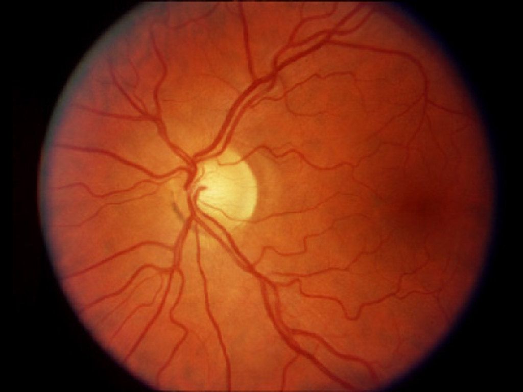
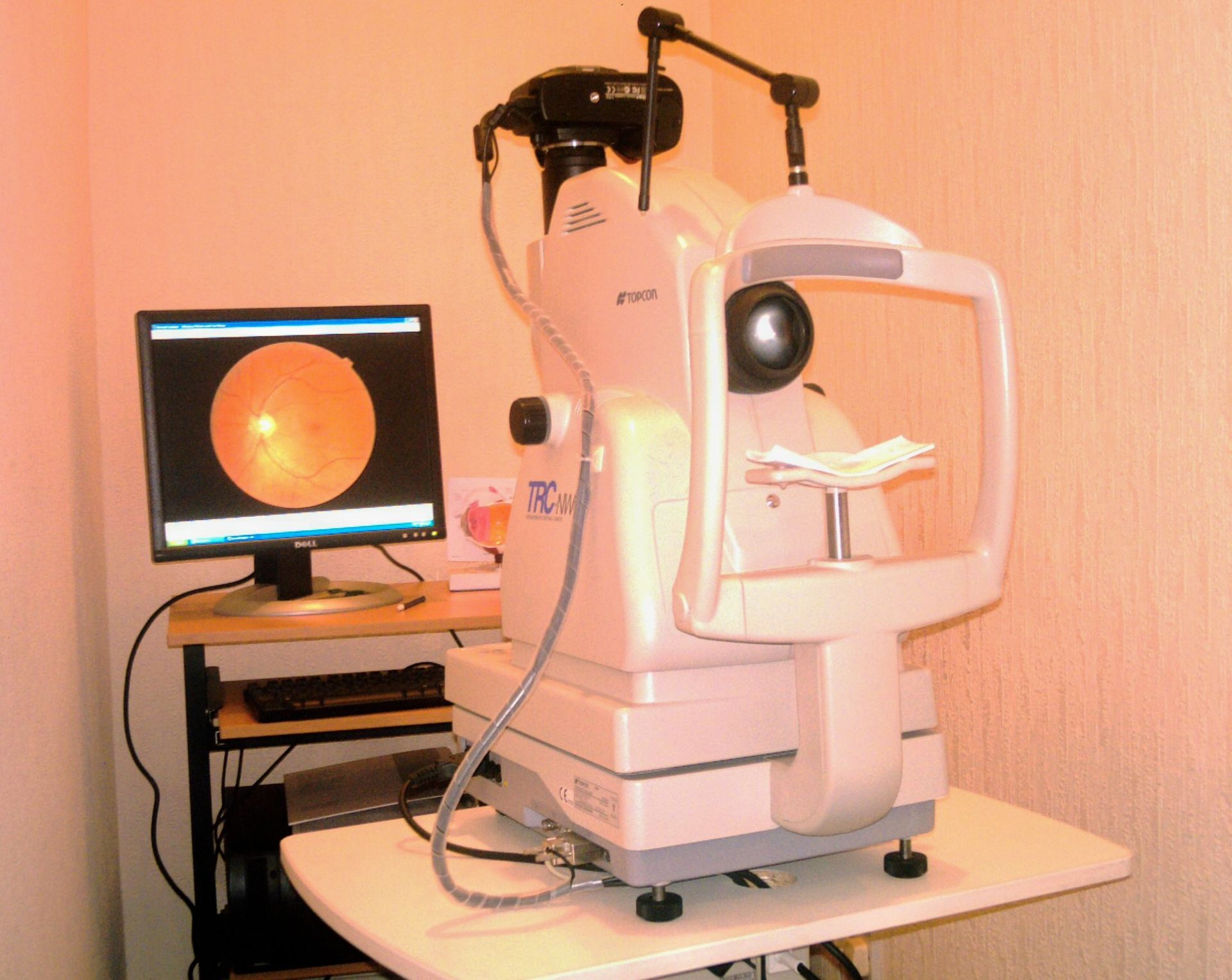
Advanced Method of Eyecare using Digital Retinal Photography.
We have installed the latest state-of-the-art digital retinal camera that allows us to offer you an enhanced type of eye health check.
The retinal camera takes high definition digital photographs of the back of each eye (the fundus), often without the need for dilation with eyedrops.
Having retinal photographs taken is painless, and provides a permanent detailed record of the fundus of each eye, which can assist in the early identification of many retinal conditions. The photographs are stored and can be compared every time you visit.
You can see the pictures of your own retinas immediately on a plasma screen in the consulting rooms. Many of our patients are fascinated when they see their own retinal photographs, and we always try to point out the main structures of the fundus. Some patients prefer not to look at the pictures...... either way, the results are invaluable